Real-Time Visualization of Autophagosome Transfer
Opening a New Era in Dynamic Decomposition Biology
For the first time in the world, the exact moment when an organelle switches its transport route-a so-called "handoff"-within a living cell has been captured in real time. This marks the first direct observation, over time, of the inter-organelle transport mechanism in cells, which until now had only been hypothesized.
The Center for Molecular Spectroscopy and Dynamics at the Institute for Basic Science (IBS) announced that it has successfully visualized, in real time and with millisecond and nanometer resolution, the moment when an autophagosome is transferred from the endoplasmic reticulum to a microtubule during autophagy.
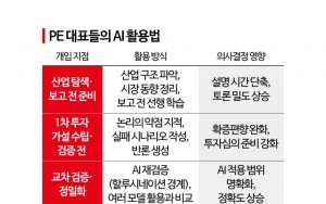
![World's First Real-Time Capture of Organelle "Handoff" in Cells... Unraveling the Mystery of Autophagosome Transport [Reading Science]](https://cphoto.asiae.co.kr/listimglink/1/2026012811265918896_1769567218.jpg) Autophagosomes, microtubules, and endoplasmic reticulum structures within living cells observed by combining fluorescence microscopy and interferometric scattering imaging. Fluorescently labeled LC3 protein was used to identify autophagosomes and the microtubule network, while interferometric scattering imaging visualized the overall cell morphology and dynamic structures of the endoplasmic reticulum. This allowed precise tracking of individual autophagosome trajectories and high-speed (200Hz) movement characteristics. Provided by the research team
Autophagosomes, microtubules, and endoplasmic reticulum structures within living cells observed by combining fluorescence microscopy and interferometric scattering imaging. Fluorescently labeled LC3 protein was used to identify autophagosomes and the microtubule network, while interferometric scattering imaging visualized the overall cell morphology and dynamic structures of the endoplasmic reticulum. This allowed precise tracking of individual autophagosome trajectories and high-speed (200Hz) movement characteristics. Provided by the research team
'A Moment Never Seen Before'... Why Was It So Difficult?
Autophagy is a core biological process in which cells maintain homeostasis by degrading and recycling damaged or unnecessary proteins and organelles. During this process, autophagosomes are formed and grow in the endoplasmic reticulum, then travel along microtubules to fuse with lysosomes.
In particular, the phenomenon in which autophagosomes switch their transport route at the junction between the endoplasmic reticulum and microtubules has long been proposed. However, because the space is extremely narrow and the movement is exceedingly fast, it had never been directly observed in living cells.
The IBS research team developed a multi-modal imaging system that combines their proprietary label-free interferometric scattering microscope, DySLIM (Dynamic Scattering-Particle Localization Interference Microscopy), with fluorescence imaging. Using this system, they tracked the position and movement of autophagosomes via fluorescence, while simultaneously observing the network structure and dynamic changes of the endoplasmic reticulum in a label-free manner.
In particular, by applying a single fluorescent label to the LC3 protein, they implemented a strategy that allows simultaneous identification of autophagosomes and microtubules using a single signal. In addition, by employing DySLIM, they achieved high-speed imaging of structural changes in the endoplasmic reticulum.
As a result, the research team succeeded in capturing, in real time, the moment when an autophagosome is transferred at the endoplasmic reticulum-microtubule junction, with millisecond temporal resolution and nanometer spatial precision.
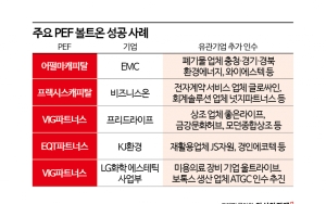
![World's First Real-Time Capture of Organelle "Handoff" in Cells... Unraveling the Mystery of Autophagosome Transport [Reading Science]](https://cphoto.asiae.co.kr/listimglink/1/2026012811291218901_1769567351.jpg) The transition process between autophagosomes and endoplasmic reticulum microtubules elucidated with millisecond time resolution and nanometer spatial precision using interference scattering microscopy. It shows the detailed movement trajectory of autophagosomes and the spatial relationship among autophagosomes, endoplasmic reticulum, and microtubules confirmed through combined fluorescence and interference scattering imaging. Provided by the research team
The transition process between autophagosomes and endoplasmic reticulum microtubules elucidated with millisecond time resolution and nanometer spatial precision using interference scattering microscopy. It shows the detailed movement trajectory of autophagosomes and the spatial relationship among autophagosomes, endoplasmic reticulum, and microtubules confirmed through combined fluorescence and interference scattering imaging. Provided by the research team
From Static Analysis to 'Dynamic Decomposition Biology'
This achievement is clearly distinguished from previous studies in that it interprets organelle transport in cells not as static snapshots, but simultaneously along both temporal and spatial axes. It is the world’s first experimental case that dynamically elucidates the inter-organelle transfer process in living cells.
Seokcheol Hong, professor of physics at Korea University, said, "By directly observing interactions among cell organelles using high-speed, high-sensitivity imaging technology, this opens a new research paradigm called dynamic decomposition biology, which interprets metabolic processes in the microscopic world across both time and space."
Minhaeng Cho, director of the IBS research group, stated, "This outcome demonstrates the potential of label-free, high-speed nano-imaging technology that does not rely on fluorescent labeling," adding, "We will further develop this into a next-generation label-free, molecule-selective imaging platform."
This research was published online on January 21 (Korean time) in the international journal ACS Nano under the title "Nanoscopy of Organelle Handoff Portals Reveals Direct Coupling between Endoplasmic Reticulum Remodeling and Microtubule-Based Transport."
© The Asia Business Daily(www.asiae.co.kr). All rights reserved.
![Clutching a Stolen Dior Bag, Saying "I Hate Being Poor but Real"... The Grotesque Con of a "Human Knockoff" [Slate]](https://cwcontent.asiae.co.kr/asiaresize/183/2026021902243444107_1771435474.jpg)


