A technology capable of precisely observing three-dimensional images of thick biological tissues such as the pancreas has been developed in Korea.
KAIST announced on the 5th that Professor Yongkeun Park's research team in the Department of Physics has developed a digital aberration correction technology that allows high-resolution observation of three-dimensional images of biological tissues without separate staining.
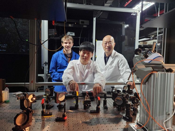 (From left) Dr. Herve Hugonnet, Integrated MS-PhD Program Student Cheolmin Oh, Professor Yonggeun Park, Department of Physics, KAIST. Provided by KAIST
Existing optical technologies showed limitations in image quality due to light scattering and optical aberrations occurring inside the tissue when observing thick biological tissues.
In contrast, by applying the technology developed by the research team, it is possible to observe thick biological tissues in real-time at high resolution through the optical memory effect. The optical memory effect is a phenomenon where scattered light tilts when the incident light tilts, enabling observation even in complex scattering media such as biological tissues.
Above all, this technology has a stronger correction effect than existing adaptive optics technology, providing the advantage of clearer observation of internal structures of biological tissues.
The research team was able to observe cellular structures inside biological tissues in more detail when applying the developed technology. They also succeeded in real-time observation of dynamic changes occurring in micrometer-sized samples.
This research result is significant in that it presents a new imaging technology that can be utilized in fields such as histopathology, new drug development, and biological research.
In particular, the research team emphasized that it is evaluated as an achievement that overcomes the limitations of deep tissue imaging that existing technologies could not surpass.
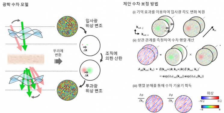 The research team succeeded in restoring high-resolution three-dimensional images with corrected aberrations by utilizing the optical memory effect, a physical property observed in light scattering. According to the team, using the optical memory effect enabled complex aberration restoration and quantitative analysis. Research flowchart. Provided by KAIST
Chulmin Oh, a combined master's and doctoral student in the Department of Physics, participated as the first author in this study, which was published online on the 17th of last month in the international journal Nature Communications. The technology developed by the research team is recognized for its potential to be widely applied in the life sciences field in the future.
Professor Yongkeun Park said, “This research is a new approach that overcomes the limitations of existing imaging technologies and will aid holography-based non-invasive biological imaging and diagnostic research. The research team plans to continue studies that enable more precise three-dimensional imaging of biological tissues to understand various life phenomena at the cellular level.”
Meanwhile, this research was conducted with support from the National Research Foundation of Korea’s Leader Research Program and the Korea Institute for Advancement of Technology’s Global Industrial Technology Cooperation Center Project.
© The Asia Business Daily(www.asiae.co.kr). All rights reserved.
![Clutching a Stolen Dior Bag, Saying "I Hate Being Poor but Real"... The Grotesque Con of a "Human Knockoff" [Slate]](https://cwcontent.asiae.co.kr/asiaresize/183/2026021902243444107_1771435474.jpg)


