A biomedical imaging technology capable of detecting risks related to stress through real-time changes in blood vessels has been developed.
On the 20th, KAIST announced that a research team led by Professor Hongki Yoo from the Department of Mechanical Engineering and Professor Jinwon Kim from the Cardiovascular Center at Korea University Guro Hospital developed a new in vivo imaging acquisition technology that compensates for vascular movement caused by heartbeats, enabling real-time observation of cellular movement within blood vessels.
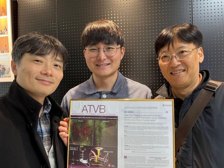 (From left) Professor Hongki Yoo, Department of Mechanical Engineering, KAIST; Minseok Jang, PhD candidate; Professor Jinwon Kim, Guro Hospital, Korea University. Courtesy of KAIST
The joint research team introduced a variable-focus lens into an in vivo optical microscope to estimate arterial movement and developed a technique to synchronize this movement with the microscope’s focal plane.
Using this technique, the team increased the inter-image correlation coefficient (a statistical indicator representing similarity between images) by four times and improved the temporal resolution (the number of images captured per unit time) by 57%, allowing real-time observation of immune cell movement within blood vessels.
In particular, by reducing image distortion caused by arterial movement and maintaining stable focus, they succeeded in real-time observation of rapidly moving immune cells inside blood vessels without missing any images.
The joint research team applied this technology to acquire in vivo images of the carotid arteries in experimental rats exposed to chronic stress and control rats, enabling quantitative evaluation of the progression of atherosclerotic lesions at the cellular resolution level.
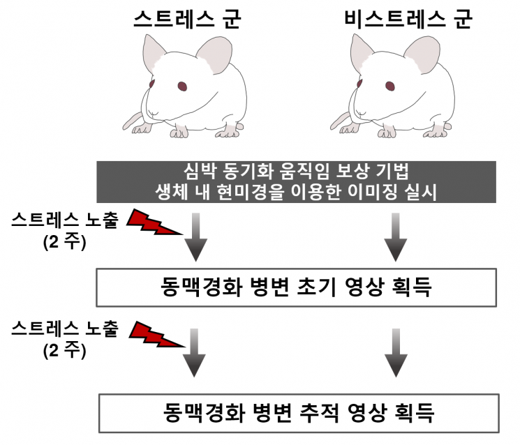
During this process, infiltration of bone marrow cells in the carotid arteries of rats exposed to chronic stress increased by 6.09 times compared to the control group, and tracking images confirmed a 2.45-fold increase in bone marrow cells.
Histological analysis also demonstrated that stress increases the size and inflammation of atherosclerotic plaques and thins the fibrous cap, thereby increasing plaque instability.
Professor Hongki Yoo of KAIST said, “This study is significant in that it enables accurate estimation of arterial movement using a non-contact method,” adding, “Above all, the joint research team ensured high survival rates of experimental animals through in vivo imaging acquisition technology, proving the effects of chronic stress through longitudinal studies.”
He further emphasized, “This technology allows real-time observation of the effects of stress on cardiovascular diseases at the cellular level with excellent temporal resolution,” and “It is expected to be an important tool for elucidating the pathogenesis of stress-related cardiovascular diseases and developing new treatments in the future.”
Meanwhile, this research was conducted with support from the National Research Foundation of Korea and the Pan-Government Medical Device Research and Development Project Group.
The study, co-first authored by Minseok Jang, a doctoral student in the Department of Mechanical Engineering at KAIST, was published online on October 10 last year in the international journal Arteriosclerosis, Thrombosis, and Vascular Biology.
© The Asia Business Daily(www.asiae.co.kr). All rights reserved.
![Clutching a Stolen Dior Bag, Saying "I Hate Being Poor but Real"... The Grotesque Con of a "Human Knockoff" [Slate]](https://cwcontent.asiae.co.kr/asiaresize/183/2026021902243444107_1771435474.jpg)


