UNIST and Kyoto University Achieve High-Resolution Imaging... Analysis of Interaction Between Mouse Sperm and Reproductive Organs
First Observation of Movement and Synchronization by Head Hook Piercing Inner Wall... Published in eLife Paper
A phenomenon in which mouse sperm move by puncturing the uterine wall with their hook-shaped heads has been captured for the first time.
A research team led by Professor Jung-Hoon Park from the Department of Biomedical Engineering at UNIST (President Jong-Rae Park), in collaboration with Professor Jae-Ik Kim from the Department of Life Sciences and Dr. Heung-Jin Ryu from Kyoto University, announced that they discovered this phenomenon by analyzing real-time videos of sperm movement inside the reproductive organs of mice.

This study was conducted to directly verify two opposing hypotheses regarding the function of the hook on rodent sperm within living tissue. Until now, the prevailing hypothesis was the ‘sperm cooperation’ hypothesis, which suggested that mouse sperm connect their hook-shaped heads like a train to increase their speed toward the egg. However, this observation did not confirm such a phenomenon.
Instead, it was observed that sperm puncture the inner walls of the uterus and fallopian tubes with the hook on their heads to move quickly. This observation supports another hypothesis, the ‘interaction hypothesis between sperm and female reproductive organs.’ The research team suggested that by puncturing the inner walls of the female reproductive organs with the hook on their heads, sperm can increase their linearity and resist strong fluid flows.
Additionally, this study observed for the first time that sperm heads align in one direction, and sperm tails synchronize their movements like synchronized swimming athletes. The research team analyzed that thanks to the stabilizing effect of the sperm hook, sperm heads are arranged and move in one direction, enabling synchronized swimming. They also proposed a new hypothesis that the hook on mouse sperm heads may be an evolutionary adaptation for such behavior.
The research team explained, “The ‘train hypothesis’ has so far only been observed in 2D culture dishes due to limitations in observation technology,” adding, “In this experiment, by observing and analyzing the actual reproductive organs, a small cluster moving in a train-like formation was found, but their movement speed was not faster than that of individual sperm.” However, they added that additional research is needed to obtain statistically significant data to completely overturn the ‘train hypothesis.’
The research team observed this phenomenon by mating genetically modified male mice whose sperm heads fluoresce green and parts of their tails fluoresce red with female mice, then extracting the reproductive organs.
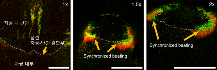
The observation was conducted using three-dimensional imaging technology based on two-photon microscopy. Two-photon microscopy is a microscope that obtains images by analyzing fluorescence emitted when two low-energy photons are irradiated onto a sample instead of one high-energy photon, preventing damage to the sample due to light energy. Also, because it uses long-wavelength light, it can observe deep inside biological tissues. The research team developed this technology by combining two-photon fluorescence phenomena with femtosecond laser-based high-speed 3D volume imaging technology.
The joint research team also secured technology to quantitatively measure sperm movement speed and characteristics through the acquired images, expecting it to aid in the development of sophisticated fallopian tube simulation chips and infertility research.
The research was supported by the National Research Foundation of Korea and the Institute for Information & Communications Technology Planning & Evaluation, and the results were published in the international life sciences journal eLife on November 22.
© The Asia Business Daily(www.asiae.co.kr). All rights reserved.
![Clutching a Stolen Dior Bag, Saying "I Hate Being Poor but Real"... The Grotesque Con of a "Human Knockoff" [Slate]](https://cwcontent.asiae.co.kr/asiaresize/183/2026021902243444107_1771435474.jpg)


