A technology capable of real-time, high-resolution observation of the dynamic changes of 'organoids' has been developed domestically. Organoids are three-dimensional mini-organs that mimic the structure and function of human organs, playing an essential role in various disease research and new drug development.
On the 14th, KAIST announced that a research team led by Professor Yongkeun Park from the Department of Physics, in collaboration with the Genome Editing Research Division of the Institute for Basic Science (IBS) and Tomocube Inc., developed an imaging technology using holotomography that enables high-resolution, real-time observation of living small intestine organoids.
 (From left) Dr. Manjae Lee, Graduate School of Medical Science, KAIST; Professor Yonggeun Park, Department of Physics, KAIST. Provided by KAIST
Organoids differ from two-dimensional cell culture methods in that they can more accurately reproduce the structure of biological tissues and cellular organization.
However, to fully understand the complex structure and dynamic biological phenomena of organoids, high-resolution real-time imaging is necessary.
Existing imaging technologies mainly require fluorescent labeling, which demands significant time investment and can cause phototoxicity and photobleaching issues. This has highlighted the need for new imaging technologies that can observe living biological samples in real time at high resolution without labeling.
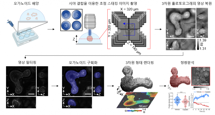
The joint research team introduced holotomography technology to address these issues, providing high-resolution images without staining such as fluorescence and enabling long-term real-time observation of dynamic changes in organoids without cellular damage.
This method allows real-time observation of three-dimensional biological samples without labels. Small intestine organoids derived from mouse small intestines were cultured in a special medium, and real-time holotomography imaging was performed and analyzed.
The technology was validated using small intestine organoids from experimental mice.
The validation involved quantitatively evaluating the growth patterns, morphological changes, and protein density of the organoids, as well as observing morphological changes after drug treatment to analyze cell death and survival rates.
As a result, the joint research team was able to capture high-resolution morphological details and dynamic activities of small organoids in vivo. It also became possible to observe the growth patterns of organoids over time and various life phenomena at the cellular level in real time.
The joint research team anticipates that this research outcome will not only open possibilities for personalized treatment using patient-derived organoids but also maximize their use as regenerative therapeutics.
Above all, the team expects this study to serve as a stepping stone that will change the paradigm of future organoid research and contribute to medical and life sciences research by enabling the understanding of various life phenomena at the cellular level through more sophisticated three-dimensional imaging.
Meanwhile, this research was conducted with support from the National Research Foundation of Korea’s Leading Research Program, KAIST research institutes, and the Institute for Basic Science. The research results were published online on the 1st in the international journal ‘Experimental & Molecular Medicine.’
© The Asia Business Daily(www.asiae.co.kr). All rights reserved.
![Clutching a Stolen Dior Bag, Saying "I Hate Being Poor but Real"... The Grotesque Con of a "Human Knockoff" [Slate]](https://cwcontent.asiae.co.kr/asiaresize/183/2026021902243444107_1771435474.jpg)


