IBS Research Team Develops 3D Hologram Microscope

[Asia Economy Reporter Kim Bong-su] A holographic microscope capable of observing brain neural networks in 3D high resolution without removing the skull has been developed.
The Institute for Basic Science (IBS) announced on the 30th that Associate Director Won-Sik Choi of the Molecular Spectroscopy and Dynamics Research Division, in collaboration with Professor Moon-Seok Kim of The Catholic University of Korea and Professor Myung-Hwan Choi of Seoul National University, developed a holographic microscope that can observe brain neural networks in 3D high resolution in living mice without removing the skull.
To observe deep inside the human body using light, it is necessary to deliver sufficient light energy and accurately measure the reflected signals. However, in biological tissues, light undergoes multiple scattering caused by collisions with various cells and aberrations that blur the image, making observation difficult.
In complex structures like biological tissues, light undergoes multiple scattering, randomly changing its propagation direction several times. During this process, the image information carried by the light is lost. Even if only a very small amount of light that has collided once with the object of interest and reflected (single-scattered wave) is selectively extracted and corrected for wavefront distortion caused by aberrations, deep observation is possible. However, multiple scattered waves interfere with this. Therefore, to obtain high-depth biological images, it is important to remove the interfering multiple scattered waves and increase the proportion of single-scattered waves.
In 2019, IBS first developed a time-resolved holographic microscope capable of removing multiple scattering and simultaneously measuring the intensity and phase of light, and observed the neural network of living fish without incision surgery. However, in the case of mice with thicker skulls than fish, severe light distortion and multiple scattering occurring in the skull made it impossible to obtain brain neural network images without removing or thinning the skull.
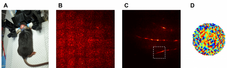
The research team developed a high-depth 3D time-resolved holographic microscope capable of observing deeper by quantifying the interaction between light and matter. They devised a method to selectively extract only single-scattered waves by utilizing the characteristic that single-scattered waves have similar reflection waveforms even when light is incident from various angles. This is a numerical calculation method analyzing the intrinsic modes of the medium (the material transmitting the wave) to find a resonant state that maximizes constructive interference (interference occurring when waves of the same phase overlap) between light wavefronts. In other words, using this method, they gathered 80 times more light on the brain neural network than before, selectively removed unnecessary signals, and increased the proportion of single-scattered waves by several tens of times.
The research team corrected wavefront distortion of light at depths previously impossible with existing technology and succeeded in obtaining high-resolution images of brain neural networks beneath the skull in mice without removing the skull, using visible light band lasers without fluorescent labeling.
The results of this study were published online on the 28th of last month in the international journal Science Advances (IF 14.136).
© The Asia Business Daily(www.asiae.co.kr). All rights reserved.
![Clutching a Stolen Dior Bag, Saying "I Hate Being Poor but Real"... The Grotesque Con of a "Human Knockoff" [Slate]](https://cwcontent.asiae.co.kr/asiaresize/183/2026021902243444107_1771435474.jpg)


