 (From left) Yesung Lee, integrated master's and doctoral student in Mechanical Robotics at GIST, Research Professor Alexander Zvanov, Professor Sung Yang.
Unlike traditional blood tests that rely on large equipment used in hospitals, a next-generation biosensor technology has been developed that can quickly and precisely check key blood indicators with only a small amount of blood.
On September 3, the Gwangju Institute of Science and Technology (GIST) announced that Professor Sung Yang’s research team from the Department of Mechanical Robotics Engineering has developed a technology that utilizes a Microfluidic Electrochemical Impedance Sensor (MEIS) to precisely analyze both the morphology and electrical properties of red blood cells simultaneously, and, based on this, calculates key indicators at the level of conventional clinical blood tests.
The Microfluidic Electrochemical Impedance Sensor is a device that measures electrochemical impedance (electrical resistance and dielectric properties) generated when a small amount of blood or cell suspension flows through a microfluidic channel equipped with electrodes. It enables non-destructive and real-time analysis. Through multi-frequency measurements, it can precisely identify a variety of biological information such as changes in red blood cell morphology and the electrical properties of cell membranes and cytoplasm. As a result, it is attracting attention as a next-generation biosensor technology for blood testing, cell analysis, and early disease diagnosis.
This technology allows for even more accurate analysis compared to existing sensors. By electrically detecting and reflecting changes in osmotic conditions as blood flows, it provides stable and highly reliable results.
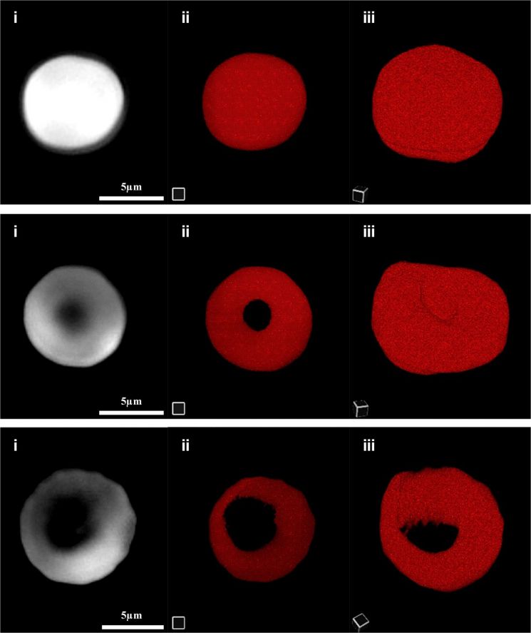 Observation of erythrocyte morphological changes under osmotic conditions using holotomography microscopy.
Blood tests are essential for the early detection of anemia, infections, cardiovascular diseases, and more, by examining various indicators such as red blood cell count, hemoglobin concentration, and plasma viscosity. However, conventional equipment requires large blood samples, expensive clinical devices, and skilled personnel, resulting in long analysis times and limitations for immediate bedside testing.
To overcome these limitations, microfluidic-based technologies capable of analyzing small amounts of blood quickly and accurately are gaining attention. Analytical methods that utilize changes in electrical signals (impedance analysis) have also attracted interest, as they allow real-time measurements without damaging cells.
The research team previously published a study that combined electrochemical impedance spectroscopy (EIS) with microfluidic channels simulating actual blood flow environments to measure the electrical properties of blood and quantitatively analyze the arrangement of red blood cells and the hydration structure around hemoglobin. Through this, they demonstrated that hematological indicators could be derived from real blood samples.
However, previous studies had limitations in precisely measuring indicators because they did not sufficiently account for the expansion or contraction of red blood cells under different osmotic conditions.
To address this, the research team observed how red blood cells change in shape and volume under different osmotic environments-such as swelling when absorbing water or shrinking when losing moisture-using optical microscopy and holotomography microscopy. They then measured the electrical responses of blood samples at various frequencies through microfluidic channels equipped with electrodes, enabling more precise detection of changes inside and outside the cells.
Based on these findings, the team calculated the dielectric properties of plasma, red blood cell membranes, and cytoplasm, and proposed a new analytical model that incorporates the contraction and expansion of red blood cells and the hydration state around hemoglobin in response to osmotic pressure changes.
As a result, the research team succeeded in calculating six major indicators used in clinical blood tests, achieving a high accuracy rate that matched more than 95% of values obtained from conventional clinical equipment.
In addition, they demonstrated that evaluating the viscosity of plasma and the internal fluid of red blood cells allows for an even more precise reflection of a patient’s health status. Notably, unlike the conventional Coulter Counter method, which only estimated cell volume at a single frequency, this study comprehensively analyzed changes in electrical signals across multiple frequencies and proposed a new analytical method that enables a comprehensive interpretation of the dielectric properties of blood components.
Professor Sung Yang stated, "This research is significant in that it has developed a technology capable of analyzing hematological indicators by incorporating changes in water content within blood. By quantifying both morphological changes and electrical properties of blood components simultaneously, it will serve as an important starting point for real-time blood testing and the development of next-generation point-of-care diagnostic devices."
© The Asia Business Daily(www.asiae.co.kr). All rights reserved.


