No Need for Expensive Lasers:
Minimally Invasive Photodynamic Therapy Also Possible
Featured on the Cover of Advanced Materials
A Korean research team has developed a technology that allows clear imaging of internal biological tissues using a common light source, such as a laser pointer, instead of an ultra-high-speed pulsed laser costing hundreds of millions of won.
On May 19, a team led by Professors Junghoon Park and Joo Jinmyung from the Department of Biomedical Engineering at UNIST announced that they have developed a nonlinear fluorescence microscopy technology that enables three-dimensional imaging of internal biological tissues using only a general continuous wave (CW) laser, by utilizing special nanoparticles.
 Professor Junghoon Park
 Professor Joo Jinmyung.
This technology achieves resolution and penetration depth comparable to those of ultra-high-speed lasers, even without such lasers. It can also be applied to photodynamic therapy, selectively stimulating only the lesion area without damaging surrounding tissues.
Biological tissues scatter light significantly, making it difficult to obtain clear internal images. For this reason, special observation techniques, such as multiphoton microscopy, are used to image biological tissues by generating fluorescence only near the focal point, thereby filtering out background noise caused by scattering. However, multiphoton microscopy requires expensive femtosecond pulsed lasers as the light source, making it challenging to use in general hospitals or laboratories.
Multiphoton microscopy suppresses background noise and enhances focus by emitting fluorescence only when two or more photons (light particles) from the laser source are simultaneously absorbed by a single molecule. With general laser sources, the probability of two photons reaching one molecule at the same time is low, so a femtosecond laser, which concentrates light and emits it in pulses to momentarily increase photon density, is necessary.
The joint research team developed a technology that can induce fluorescence only at the focal point without a femtosecond pulsed laser, by using upconversion nanoparticles (UCNPs).
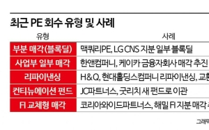
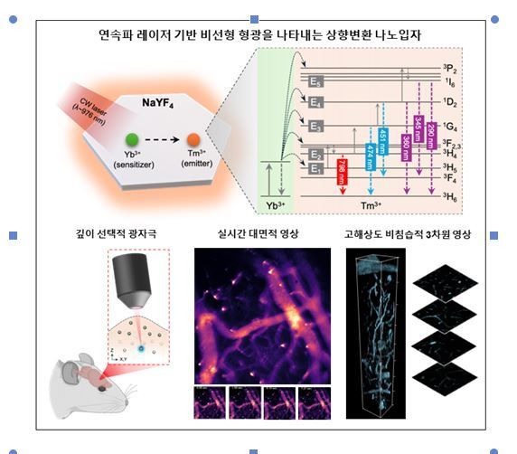 Principle and Application Examples of Nonlinear Fluorescence Imaging Using Continuous Wave Laser-Based Upconversion Nanoparticles.
After injecting the nanoparticles into the biological site via the bloodstream and irradiating them with a general continuous wave laser, the nanoparticles absorb photons from the laser one by one, accumulate energy, and emit it as ultraviolet or blue fluorescence. The emission intensity increases nonlinearly, such as by the square or cube of the light intensity, so strong fluorescence appears only in regions where light is concentrated, such as near the focal point.
Using this technology, the research team successfully imaged the cerebral blood vessels of a living mouse at a depth of about 800 micrometers with high resolution. This is about six times deeper than what is possible with confocal microscopy and is comparable to the penetration depth of multiphoton fluorescence microscopy. In wide-field mode, which enables rapid imaging of large areas, the flow of blood could be observed in real time at 30 frames per second.
The developed technology can also reduce side effects in photodynamic therapy (PDT) where tissues other than the lesion are damaged. In photodynamic therapy, light is used to penetrate and destroy the lesion, but normal tissues along the path of the light can also be damaged in the process.
By utilizing the principle of generating fluorescence only near the focal point, it is possible to selectively stimulate only the lesion area, enabling precise photostimulation therapy that does not affect surrounding tissues. The research team also succeeded in experiments that activated ultraviolet-reactive substances at specific depths using ultraviolet light emitted by the upconversion nanoparticles.
This research was led by Dr. Jungmo Kim and Dr. Seunghoon Lee of UNIST as the first authors.
The joint research team stated, "This is a technology that enables both high-resolution bioimaging and precise phototherapy without expensive ultra-high-speed lasers," and added, "If used in conjunction with conventional diagnostic equipment such as MRI, it will also help precisely track cerebral blood flow or localized metabolic responses in medical settings."
The research was supported by the National Research Foundation of Korea under the Ministry of Science and ICT, the Institute for Information & Communications Technology Planning & Evaluation, the Korea Dementia Research Center, the Korea Regenerative Medicine Project Group, and the POSCO Cheongam Foundation. The results were published as a cover paper in the world-renowned journal 'Advanced Materials' in the field of materials science on May 12.
© The Asia Business Daily(www.asiae.co.kr). All rights reserved.


