An analytical method has been developed to safely develop exosome-based therapeutics, which are considered promising new drug candidates. Exosomes are small vesicles composed of nano-sized double lipid bilayers secreted by cells.
The Korea Research Foundation announced on the 5th that a joint research team led by Dr. Youngwoo Cho and Dr. Youngwook Noh from Osong Advanced Medical Industry Promotion Foundation, together with Dr. Hyesun Park and researcher Miyoung Cho from the Korea Basic Science Institute, developed a quantitative analysis method capable of evaluating the in vivo distribution of exosomes.
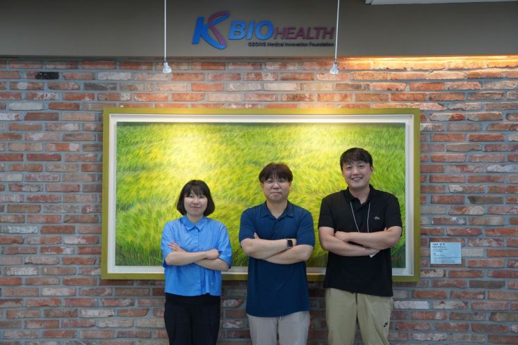 (From left) Jomiyoung, Researcher at Korea Basic Science Institute, Dr. Youngwook Noh and Youngwoo Jo, Osong Advanced Medical Industry Promotion Foundation. Provided by National Research Foundation of Korea
In vivo distribution evaluation is one of the test items required for clinical trial approval of new drugs. It is particularly significant because analyzing the in vivo movement, distribution, and persistence using animal models can determine the extent of both targeted and off-target effects of therapeutics.
Exosome-based therapeutics are advanced biopharmaceuticals developed by isolating and purifying extracellular vesicles secreted from living cells. Development is actively underway for therapeutics, disease diagnostic tools, and drug delivery systems.
However, due to the lack of clear analytical methods, there are global difficulties in entering clinical trials. To date, no exosome-based therapeutic has been commercially approved.
For in vivo distribution evaluation of exosomes, imaging analysis based on labeling methods using lipophilic dyes and radioisotopes is mainly utilized. However, these labeling substances alter the natural and biological properties of exosomes, leading to limitations in analysis.
Considering this, the joint research team focused on accurately measuring the in vivo distribution of exosomes through proteins and RNA while maintaining their biological characteristics.
First, they confirmed the presence of mitochondrial DNA in exosomes and set it as the analysis target. This means that when exosomes derived from human cells are administered to experimental animals, the analysis target can be clearly distinguished.
Mitochondrial DNA refers to DNA present in the mitochondria of the cytoplasm and varies by animal species, making it a reliable tool for species identification.
Following this testing method, the research team detected unmodified exosomes and analyzed them using quantitative PCR (a molecular biology technique that replicates and amplifies desired DNA segments to estimate the amount of specific DNA sequences). They confirmed that the amount of mitochondrial DNA varies among exosomes isolated from different cells.
In the in vivo distribution evaluation of exosomes administered via the tail vein of rodents, they verified that the exosomes were widely distributed across all organs after administration. They also compared and analyzed the existing imaging analysis with the quantitative PCR method to validate the testing method.
Researcher Youngwoo Cho stated, “This study can provide important information for the development of exosome-based therapeutics because it allows accurate evaluation of the in vivo distribution of unmodified exosomes.” He added, “It is expected to establish the scientific basis necessary to expedite clinical approval of exosome therapeutics.”
Meanwhile, this research was conducted with support from the Bio and Medical Technology Development Project promoted by the Ministry of Science and ICT and the Korea Research Foundation. The research results were published on July 17 in the international journal in the field of extracellular vesicle research, the Journal of Extracellular Vesicles.
© The Asia Business Daily(www.asiae.co.kr). All rights reserved.


