Latest Issue of the Journal of the Korean Medical Association Released
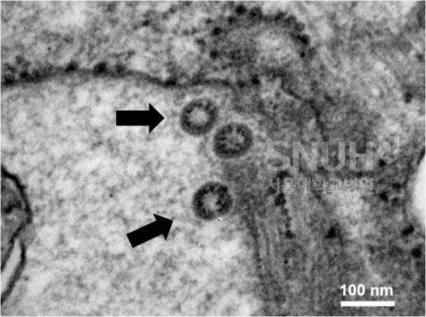 The research team led by Professors Park Wan-beom and Oh Myung-don from the Department of Infectious Diseases at Seoul National University Hospital released electron microscope images on the 19th that allow the COVID-19 virus to be seen with the naked eye.
 [Photo by the Department of Infectious Diseases, Seoul National University Hospital / Image source: Yonhap News]
[Asia Economy Reporter Kim Heung-soon] Electron microscope images that allow the novel coronavirus (COVID-19) to be visually confirmed have been revealed for the first time in South Korea.
According to the latest issue of the Journal of the Korean Medical Science (JKMS) on the 19th, a research team led by Professors Park Wan-beom and Oh Myung-don from the Department of Infectious Diseases at Seoul National University Hospital successfully isolated and cultured the virus from the first confirmed domestic patient (born in 1984, female of Chinese nationality) who entered from Wuhan, Hubei Province, China, and was diagnosed positive on the 20th of last month, and captured images using an electron microscope.
In their paper, the researchers stated, "The virus shows 99.7% similarity compared to the COVID-19 strain that originated in China, but there are nine genetic mutations," and added, "Further studies are needed to understand the academic significance of these genetic mutations." The electron microscope magnified images reveal the spikes surrounding the virus particles. The term 'corona' comes from Latin, meaning crown. The coronavirus is named for the crown-like spikes on its surface.
© The Asia Business Daily(www.asiae.co.kr). All rights reserved.


