40s Patient with Brain Lesions from Parasite Infection
Drank Contaminated Pond Water and Ate Raw Fish
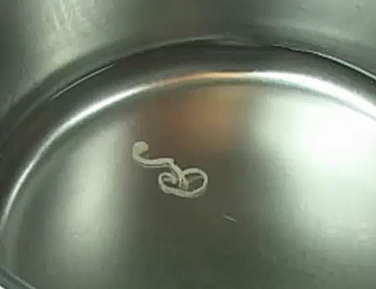
Korean Medical Team Successfully Removes Lesions via Surgery
Domestic researchers successfully removed a living parasite from the brain of a patient in their 40s.
On the 30th, a joint research team led by Professor Baek Seon-ha of the Department of Neurosurgery at Seoul National University Hospital and Professor Park Hye-ran of the Department of Neurosurgery at Soonchunhyang University Seoul Hospital announced that they diagnosed and treated a patient in their 40s who developed brain lesions due to Sparganosis parasite infection, and reported it to the academic community.
Sparganosis is a rare parasitic infection disease caused by larvae penetrating the body and migrating to the brain through the bloodstream. It can mainly occur when drinking contaminated water or consuming raw or undercooked wild animal meat or fish. When the infected parasite moves to the brain, symptoms such as headaches and vomiting appear, and over time, it can lead to neurological problems such as seizures, visual field defects, and sensory abnormalities. Additionally, if the parasite reaches around the orbit, the eyes and surrounding areas become itchy, painful, swollen, and tear excessively. Although antiparasitic drugs can be administered to alleviate symptoms, there is a very high risk of recurrence afterward, so surgical removal of the parasite is considered the only treatment method.
Initially, the patient visited Seoul National University Hospital complaining of severe headaches and vomiting. At that time, magnetic resonance imaging (MRI) showed a lesion in the left posterior part of the brain, which was suspected to be a brain tumor. The medical staff recommended surgery, but the patient refused treatment and was discharged as symptoms improved. Seven months later, the patient returned to the hospital with severe headaches and generalized seizures. Follow-up MRI at that time showed that the lesion had moved from the left occipital lobe to the left parietal lobe near the top of the left side of the brain, raising suspicion that it was not a simple brain tumor. Furthermore, it was found that the patient had previously drunk contaminated pond water and consumed raw fish and undercooked wild animal meat.
Considering various circumstances, the medical team suspected a parasitic infection and conducted various tests, diagnosing the symptoms as Sparganosis. They performed a craniotomy to open the patient's skull and removed the living Sparganosis larvae from the brain. Professor Baek said, "When lesions move in imaging tests, the possibility of parasitic infection must be considered," and added, "Avoiding contaminated water and thoroughly cooking wild animal meat or fish, as well as following personal hygiene rules, are key to prevention." The research results were published in the recent issue of 'Neurology,' the journal of the American Academy of Neurology.
© The Asia Business Daily(www.asiae.co.kr). All rights reserved.
![Clutching a Stolen Dior Bag, Saying "I Hate Being Poor but Real"... The Grotesque Con of a "Human Knockoff" [Slate]](https://cwcontent.asiae.co.kr/asiaresize/183/2026021902243444107_1771435474.jpg)


