A study has found that adolescents with depression who respond to antidepressant treatment are more likely to experience recovery of atrophied nerve cells and maintain a relaxed brain state.
A research team led by Professor Kim Jaewon from the Department of Child Psychiatry at Seoul National University Children's Hospital analyzed neurobiological changes occurring in the brain during the treatment of depressive symptoms in 95 adolescents aged 12 to 17 who were treated with antidepressants, producing these results.
Generally, antidepressants are used to treat depression in adolescents. Selective Serotonin Reuptake Inhibitors (SSRIs) are prescribed as the first-line pharmacological treatment for adolescents exhibiting moderate or more severe depression.
However, SSRIs as a single agent have a remission rate (response rate to medication) of 55%, and when combined with other treatments such as cognitive behavioral therapy, the remission rate is 60%. To improve the treatment efficacy of adolescent depression, it is important to understand the neurobiological changes related to symptom improvement and to identify treatment markers.
Nevertheless, research on the neurobiological treatment mechanisms of adolescent depression remains insufficient worldwide. From 2015 to 2018, the research team analyzed how SSRI treatment affects the neurobiological characteristics of the brain in 95 adolescents with depression and 57 adolescents without depression who visited the 'Mood and Anxiety Clinic' at Seoul National University Children's Hospital. The depressed adolescents were treated with escitalopram, an SSRI antidepressant, for eight weeks.
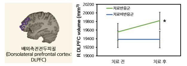
The research team classified adolescents whose depressive symptoms decreased by more than 40% after treatment compared to before treatment as the treatment response group, and those who did not meet this criterion as the non-response group. They then analyzed the correlation between changes in depressive symptoms and changes in the volume and resting-state functional connectivity (rsFC) of the dorsolateral prefrontal cortex (DLPFC). The DLPFC is one of the major brain regions involved in emotional regulation and cognitive control.
 Professor Kim Jaewon, Department of Child Psychiatry, Seoul National University Children's Hospital.
Analysis results showed that about 54% of adolescents with depression were classified as the treatment response group after antidepressant treatment. The treatment response group showed an increase in the volume of the dorsolateral prefrontal cortex compared to the non-response group.
It is known that the reduction in prefrontal cortex volume observed in depression is related to neural atrophy. The increase in dorsolateral prefrontal cortex volume after antidepressant treatment suggests the possibility of recovery of nerve cells that had atrophied due to depression, the research team explained.
Professor Kim Jaewon stated, "This study is an important result showing that antidepressant treatment in adolescent depression can be accompanied by changes in brain structure and functional connectivity responsible for emotional regulation and cognitive control," adding, "Data on changes in brain structure and functional connectivity may be utilized as biomarkers for antidepressant treatment in the future."
© The Asia Business Daily(www.asiae.co.kr). All rights reserved.


