IBS Research Team Develops High-Speed Imaging Technology
Domestic researchers have developed a high-speed scanning technology that creates brain connectivity maps by projecting images onto the brain like a movie projector. This technology is expected to be utilized in the development of treatments for degenerative brain diseases such as dementia and drug addiction.
The Institute for Basic Science (IBS) announced on the 31st that the research team led by Kim Sung-ki, head of the Brain Science Imaging Research Center, developed a technology that freely controls the cerebral cortex activity of living mice using optogenetic stimulation patterned by a beam projector, and scans the entire brain area with functional magnetic resonance imaging (fMRI) to create brain connectivity maps. The research team of Professor Choi Myung-hwan from the Department of Biological Sciences at Seoul National University also participated in this study.
Brain function is determined by interactions between different brain regions. Therefore, understanding the connectivity of each brain region is the starting point for brain function research. Optogenetics literally combines ‘light (Opto)’ and ‘genetics (Genetics).’ It is a technique that uses genetic methods to express light-sensitive proteins in specific cells and controls these cells using light. It is widely used to study causal relationships between single brain regions, whole brain circuits, and behavior by regulating the activity of brain cells in specific areas. fMRI scans the entire brain activity and can confirm changes throughout the brain caused by optogenetically induced activity in specific brain regions.
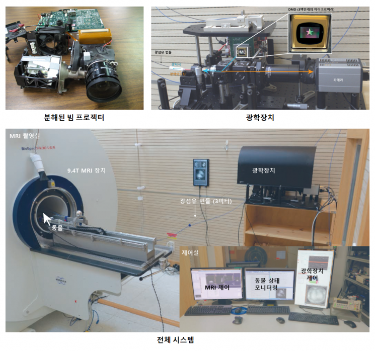
Until now, optogenetic fMRI involved invasive surgery to insert optical fibers into target brain regions and stimulate them by delivering light through these fibers. However, this method can cause unexpected damage to the brain. There is also a limitation in that many optical fibers cannot be inserted into a single experimental subject. Therefore, to study numerous brain regions, a large number of experimental animals were inevitably required. Additionally, controlling differences between experimental subjects to study connectivity differences between brain regions was challenging.
The research team devised a method to directly project light onto the cerebral cortex of mice instead of inserting optical fibers. To solve the problem of light not penetrating due to the mouse’s skull, they thinned the skull using a dental drill and treated it with chemicals to create a thinned-skull cranial window that allows the brain to be viewed. Using a beam projector equipped with a high-power optogenetic stimulation laser, they directly projected light over the entire cerebral cortex to enable effective optogenetic stimulation.
The optical stimulation via the beam projector is not a simple point but projects light onto the brain surface like screening a movie. In other words, it can freely generate desired patterns in desired brain regions. This makes it possible to deliver optical stimulation to multiple regions freely in a single experiment, rather than a fixed single region.
Using this technology, the research team sequentially stimulated various brain regions across the cerebral cortex in a single mouse experimental subject. Then, by scanning the entire brain with fMRI and analyzing the large amount of data on brain activation responses obtained, they were able to complete an effective connectivity map that shows the causal relationships of interactions between brain regions.
They anesthetized mice with two commonly used anesthetics in animal research, Isoflurane and Ketamine-Xylazine, respectively, and compared and analyzed the effective connectivity of the mouse brain under different anesthesia conditions. As a result, they found that Isoflurane selectively weakened connectivity between specific cerebral cortex regions and midbrain areas. This demonstrates that optogenetic fMRI using patterned stimulation can effectively measure changes in effective connectivity induced by specific brain states.
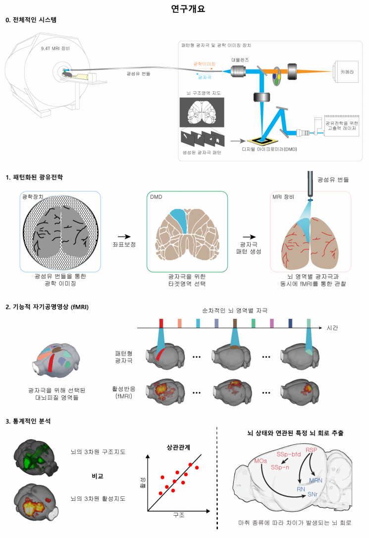
This study presented a new method to extract brain circuits associated with specific brain states, which is expected to play an important role in elucidating the mechanisms of brain function decline caused by brain diseases and drugs.
Director Kim said, “This technology is an innovative method to map functional circuits from various cerebral cortex regions to the whole brain in living animals and measure changes according to brain states,” adding, “It has become possible to conduct in-depth research on brain structure and function by directly comparing effective connectivity and structural connectivity of the entire brain.” He further explained, “In the future, it can be used to study the relationship between altered brain function and specific brain circuits caused by drugs, diseases, or physiological factors,” and “It is expected to contribute to elucidating the neurophysiological mechanisms of brain diseases and drug addiction and to developing treatment technologies.”
The research results were published online on the same day in the international neuroscience journal Neuron.
© The Asia Business Daily(www.asiae.co.kr). All rights reserved.
![From Bar Hostess to Organ Seller to High Society... The Grotesque Con of a "Human Counterfeit" [Slate]](https://cwcontent.asiae.co.kr/asiaresize/183/2026021902243444107_1771435474.jpg)


