IBS Joint Research Team
[Asia Economy Reporter Kim Bong-su] An ultra-fine endoscope thinner than a syringe needle, capable of observing capillaries and the nervous system in 3D, has been developed.
The Institute for Basic Science (IBS) announced on the 2nd that Choi Won-sik, Deputy Director of the Molecular Spectroscopy and Dynamics Research Group (Director Cho Min-haeng) and professor at Korea University, in collaboration with Professor Choi Young-woon of Korea University, developed an endoscope technology thinner than a syringe needle and succeeded in obtaining three-dimensional images of biological structures smaller than bacteria.
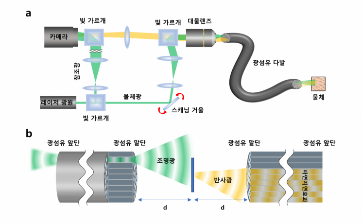
An endoscope is an imaging device designed to capture images inside narrow spaces or within the human body. Generally, an endoscope inserts a slender imaging device into the object to be observed to receive signals; a camera is attached to the probe tip for direct observation, or images are obtained using optical fibers that transmit information via light. When using a camera sensor, the probe thickness increases, sometimes requiring a small incision in the skin for insertion. In contrast, endoscopes using bundles of optical fibers can be made thinner, minimizing incision size and patient discomfort.
However, conventional fiber optic endoscopes have difficulty producing clear images due to empty spaces between the cores (the material inside the optical fiber that transmits light) of individual fibers. Additionally, self-reflection at the fiber bundle’s end interferes with observing only the desired signals, making it difficult to observe biological structures with low reflectivity. Fluorescent staining is required for observation, but its application to the human body is limited.
The research team overcame the limitations of existing fiber optic endoscopes by developing an ultra-thin endoscope capable of high-resolution observation without attaching lenses or any equipment to the fiber bundle’s end. The team focused light into one fiber within the bundle to illuminate an object located at a certain distance from the fiber. Light reflected from the object transmitted information about the object through several other fibers. They measured the reflected holographic images obtained and corrected distortions occurring in each core to acquire high-resolution images.
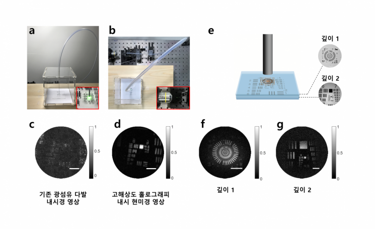
The developed endoscope does not have any equipment attached to the fiber tip, resulting in a probe diameter of 350μm (micrometers), which is thinner than a typical syringe needle placed on the skin (approximately 500μm). Using this, they obtained image information without fluorescent staining even from biological samples with very low reflectivity, such as the villi in a mouse’s small intestine.
Notably, the newly developed endoscope can capture microscope-level high-resolution images that conventional fiber bundle endoscopes cannot achieve. It can distinguish objects separated by about 850nm (nanometers). For reference, bacteria are approximately 1,000nm (1μm, or one-millionth of a meter) in size. By correcting the measured holographic information, multi-depth 3D images can also be reconstructed, distinguishing objects separated by about 14μm in depth.
Deputy Director Choi Won-sik said, “We have developed a revolutionary ultra-thin, high-resolution endoscope,” adding, “This opens the possibility of early diagnosis of diseases with minimal skin incisions in areas difficult to access with conventional endoscopes, such as the lungs, capillaries, and even the brain’s nervous system.”
The research results were published online on the 2nd of last month in the international journal Nature Communications (IF 17.69).
© The Asia Business Daily(www.asiae.co.kr). All rights reserved.


