Gwangju Institute of Science and Technology Professor Jeong Eheon’s Team Develops Quantitative Brain Blood Flow Measurement System
Analyzes Interference Patterns Created by Laser Beam Irradiation to Measure Brain Blood Flow Changes and Velocity
[Asia Economy Reporter Kim Bong-su] An optical imaging technology capable of quantitatively measuring changes and speed of cerebral blood flow has been developed by a domestic research team. It is expected to present new treatment methods for vascular diseases such as stroke.
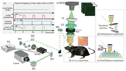
The Gwangju Institute of Science and Technology (GIST) announced on the 23rd that a team led by Professor Jeong Ui-heon of the Department of Biomedical Engineering succeeded in developing a system that can quantitatively measure changes and speed of cerebral blood flow when cerebral infarction occurs on the surface of the brain by analyzing interference patterns (speckles) generated by irradiating laser light onto the brain.
The human brain accounts for only 2% of body mass but consumes 20% of the body's oxygen and nutrients. Blood flow increases in areas where brain neurons are activated to smoothly supply oxygen and glucose. Therefore, measuring changes and speed of cerebral blood flow is very important for understanding cerebral metabolism and cerebrovascular pathology.
Existing research methods could observe changes before and after blood flow movement but had limitations in measuring the speed of blood flow. The research team developed a method to quantitatively measure the speed of dynamically changing blood flow in real time using only speckle analysis without mathematical modeling or calibration. Through this, they were able to objectively compare the efficacy of new treatments for vascular diseases by showing real-time changes in blood flow as quantitative velocity maps in a preclinical ischemic stroke model.
In particular, they utilized the principle that the real-time movement of blood cells inside blood vessels in living tissues is reflected in laser speckles. By viewing speckles as a type of particle and analyzing spatiotemporal changes, they succeeded in quantitatively deriving the actual velocity field. This required an extremely short camera exposure time, which the research team implemented using an acousto-optic modulator, enabling application to animal disease models.
Professor Jeong said, "This study overcame the limitations of existing laser speckle imaging and proposed a methodology to quantitatively analyze blood flow velocity in living tissues," adding, "It is expected to be applied to the development of stroke treatments based on animal models and clinical research on vascular diseases in the future."
The research results were published online on the 13th in the optical science journal ‘Optica’.
© The Asia Business Daily(www.asiae.co.kr). All rights reserved.


