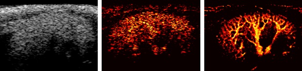
 The photo on the left is a conventional ultrasound image, and the middle one is an image captured using the existing contrast-enhanced ultrasound Doppler vascular imaging technique with a contrast agent. The photo on the right was taken using the newly developed super-resolution ultrasound imaging technique. While the conventional ultrasound imaging technique could not observe microvascular structures, the developed super-resolution ultrasound imaging technique allows for the observation of fine microvascular structures measuring only tens of micrometers.
[Asia Economy Reporter Hwang Junho] Domestic researchers have developed a technology that enhances the resolution of ultrasound images by up to 8 times. This overcomes the acoustic diffraction limit, which was considered the spatial resolution limit of ultrasound imaging. It is evaluated as a technology that can play a crucial role in various disease treatments, such as confirming the rupture vulnerability of plaques caused by atherosclerosis or monitoring real-time blood flow changes in the brain.
The research team led by Professor Yoo Jaeseok from the Department of Robotics Engineering at Daegu Gyeongbuk Institute of Science and Technology (DGIST) and a research team from the University of Pittsburgh School of Medicine in the United States announced on the 2nd that they developed super-resolution ultrasound imaging technology, and their related research results were published in Kidney International, an international journal in the field of nephrology.
The research team succeeded in implementing images with a resolution improved by more than 4 to 5 times by using localization technology that distinguishes and locates individual signals of ultrasound contrast agents. By utilizing this technology, it is possible to observe microvessels as small as 32 micrometers. Previously, only microvessels about 150 to 200 micrometers in size could be observed.
The research team also increased the image processing speed. By applying deconvolution, a signal processing technique used in astronomy, they reduced the data collection period by nearly 150 times compared to before. This made it possible to process images within less than one second. Using this technology, the team succeeded in observing the progression from acute kidney injury to chronic kidney disease, which was previously unobservable with conventional ultrasound.
Professor Yoo Jaeseok of the Department of Robotics Engineering said, "The technology developed this time has proven its effectiveness by observing the progression of diseases that were impossible to diagnose with existing ultrasound imaging devices," adding, "We are currently researching technology to implement super-resolution images in 3D, and we will develop it into a technology that can be used in actual clinical practice in the future."
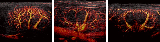 This is the result of monitoring kidneys with acute kidney injury using a super-resolution ultrasound imaging technique.
© The Asia Business Daily(www.asiae.co.kr). All rights reserved.
![Clutching a Stolen Dior Bag, Saying "I Hate Being Poor but Real"... The Grotesque Con of a "Human Knockoff" [Slate]](https://cwcontent.asiae.co.kr/asiaresize/183/2026021902243444107_1771435474.jpg)


