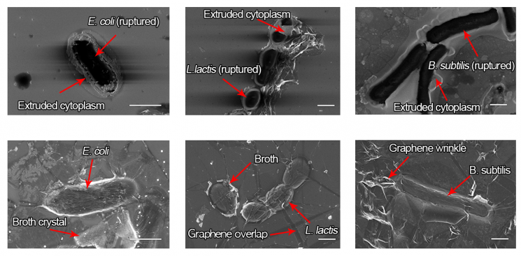
 These are scanning electron microscope images of dead cells observed using conventional electron microscopy technology (top) and living cells observed using a graphene liquid cell (bottom).
[Asia Economy Reporter Junho Hwang] A technology that allows real-time observation of the process by which living bacteria or viruses infect our body's cells has been developed domestically. By utilizing this technology, the infection process of infectious disease viruses such as the novel coronavirus infection (COVID-19) can also be observed. It is expected to open a path to visually confirm whether COVID-19 treatments are actually effective.
Real-time Observation of Living Cells' Movements
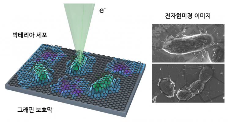 This is a schematic diagram of the cell observation method using the graphene liquid cell employed in this study, along with scanning electron microscope images of living cells observed using this method.
A joint research team led by Professor Yuk Jong-min of the Department of Materials Science and Engineering at the Korea Advanced Institute of Science and Technology (KAIST) and Professor Han Young-gi of the ITA Graduate School of Convergence at Kyungpook National University announced on the 26th that they succeeded in observing living cells in real time using an electron microscope, and their related paper was recently introduced in the international journal Nano Letters.
This research is significant in that it proposed a method to observe living cells in real time. Typically, electron microscopes are used for cell observation, but since electrons have energy thousands of times higher than visible light, structural damage to cells is inevitable during observation. Accordingly, the cryo-electron microscopy technique, which won the 2017 Nobel Prize in Chemistry, is used, but it requires fixing and stabilizing the observation target. This means it is difficult to capture the movements of living cells.
The research team solved this problem by using graphene, an atomic film 0.2 nanometers thick (1 nanometer is one hundred thousandth the thickness of a human hair) separated from graphite with a two-dimensional layered structure.
The team confirmed that by using graphene to encapsulate cells and liquids, structural changes in cells caused by dehydration inside the high vacuum of the electron microscope can be prevented. Graphene exhibits an effect of decomposing reactive oxygen species that become more aggressive due to the electron beam, revealing that cells covered with graphene do not lose activity even when exposed to electrons 100 times stronger than cells not covered with graphene.
Contributing to COVID-19 Treatment
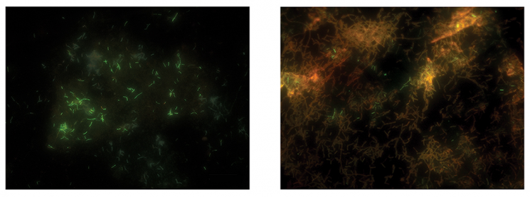 Verification of cell viability through fluorescence analysis after observing the sample using a graphene liquid cell (left) and a conventional electron microscope (right).
Observing the actual mechanisms of various cells means being able to see various biological responses in real time.
Professor Yuk stated, "The results of this research can be expanded to real-time electron microscopy observation of proteins or DNA smaller than cells, and it is expected to fundamentally elucidate various biological phenomena in the future."
This technology can also contribute to the development of COVID-19 treatments. Professor Yuk explained, "By utilizing this technology, viruses can also be observed. Once therapeutic substances are developed, it will be possible to visually confirm how the infection by viruses is blocked and the efficacy of the treatment."
© The Asia Business Daily(www.asiae.co.kr). All rights reserved.


